前回からの続きです。症例4の解説(その6) | 英語好きな歯科医の診療日記 (hn-dentist-english-blog.com) 症例の全体像はこちらです。症例4(上顎欠損部をサイナスリフト後インプラントで修復した全顎治療のケース) | 英語好きな歯科医の診療日記 (hn-dentist-english-blog.com)
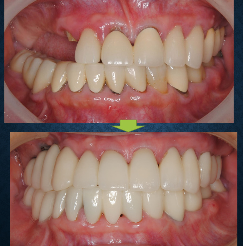
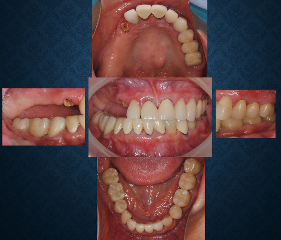
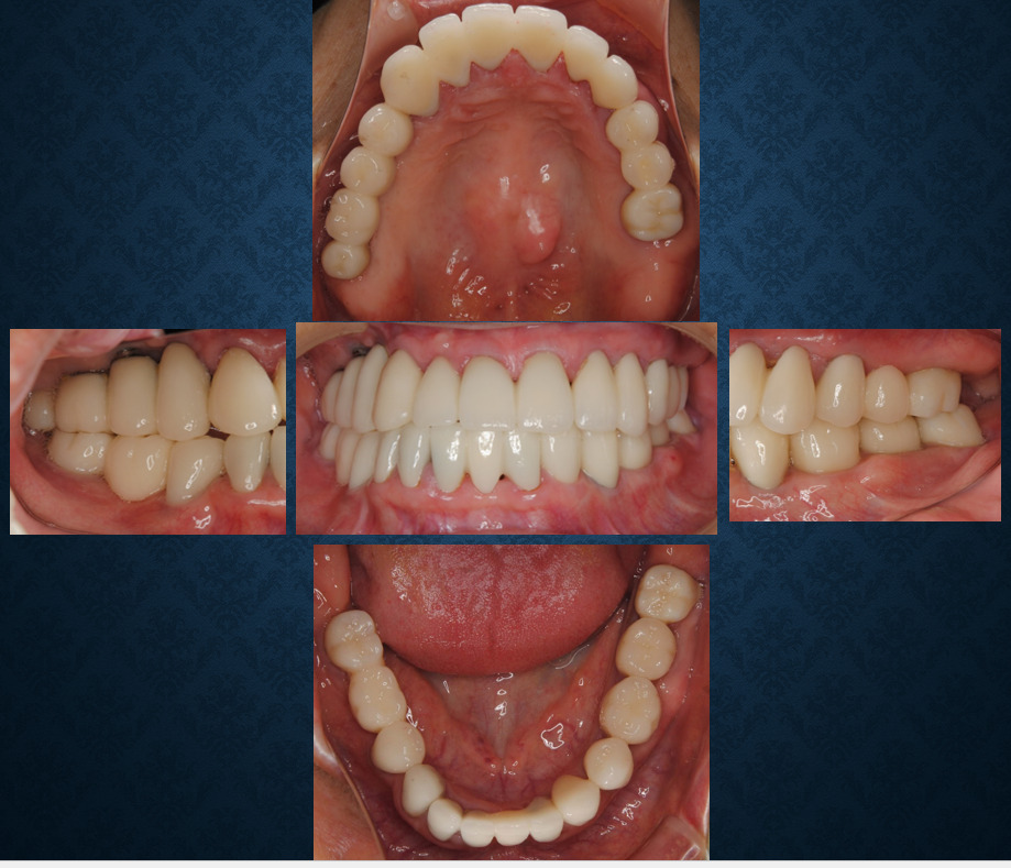
術前後の正面観です。最終的な被せ物はオールセラミック(ジルコニア)です。
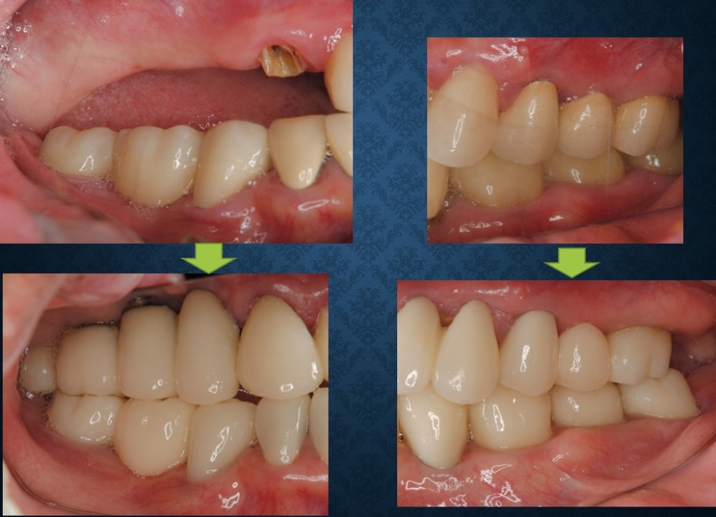
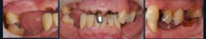
術前後の左右側面観です。
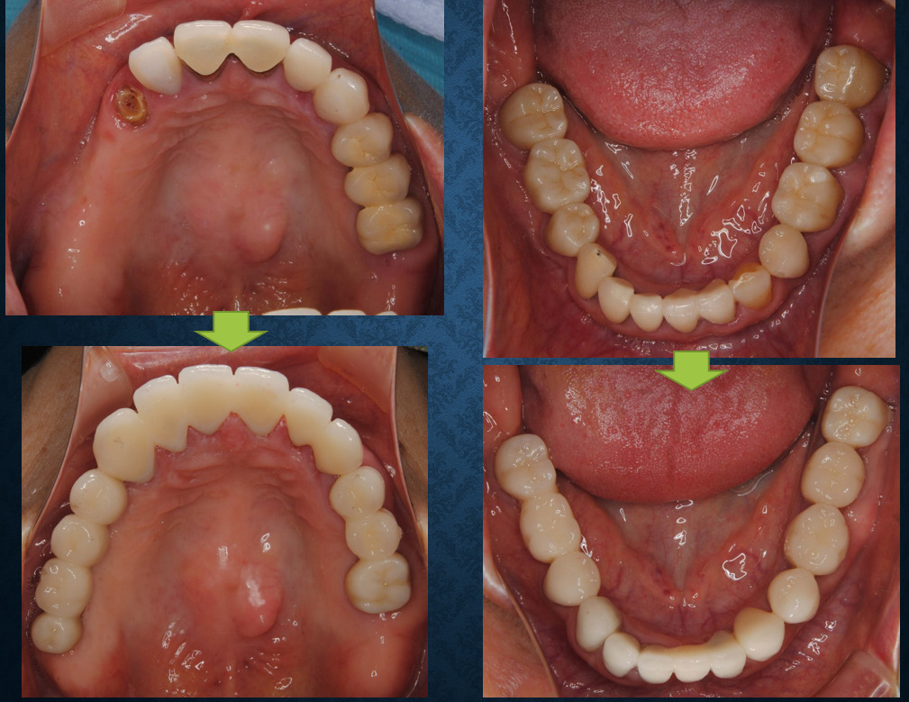
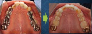
術前後の上下咬合面観です。
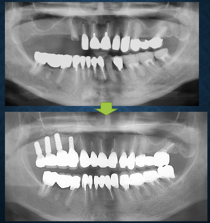
術前後のレントゲンです。
最初に比べれば咬合平面の左右の傾きの差が改善されました。
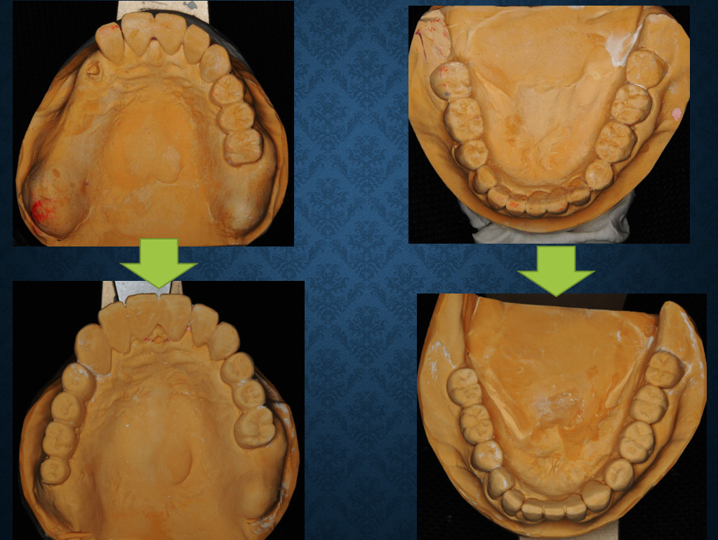
術前後の模型上下咬合面観です。
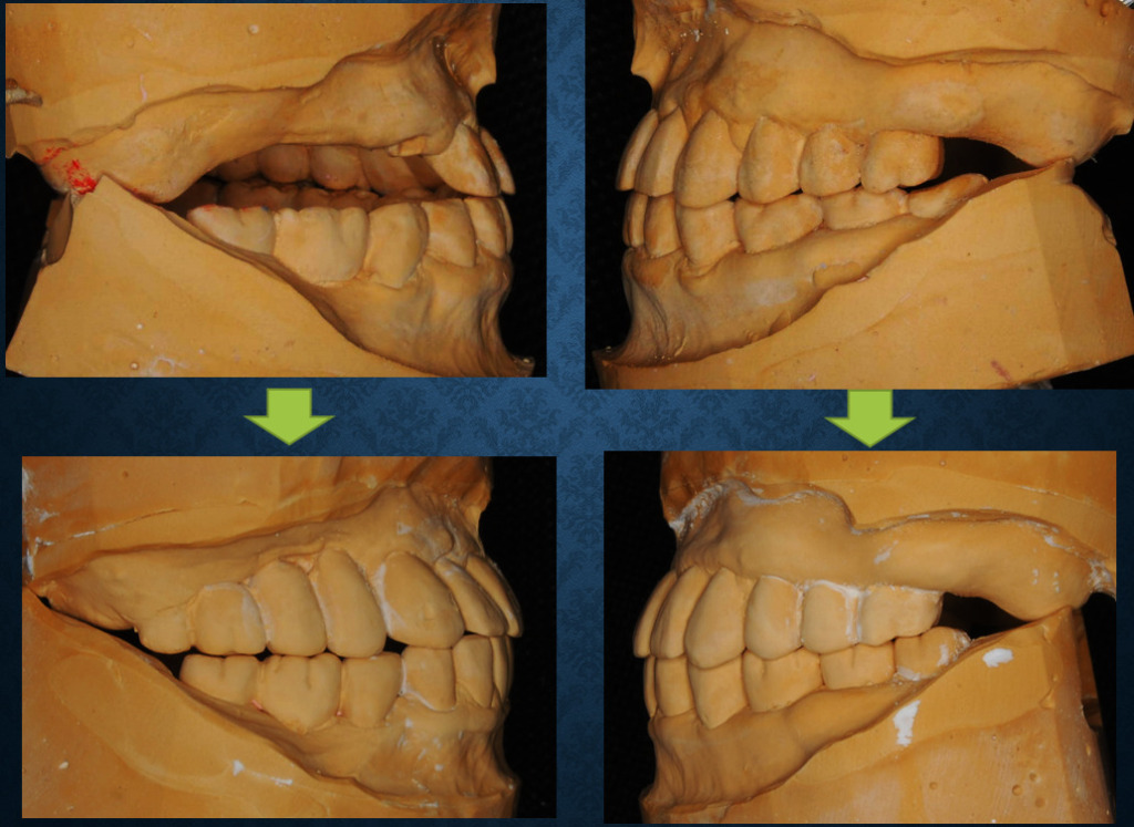
術前後の模型左右側面観です。
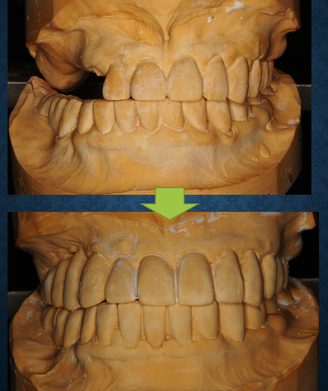
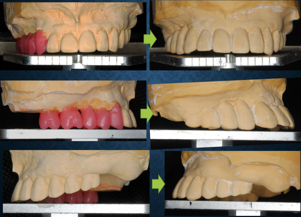
術前後の模型正面観です。
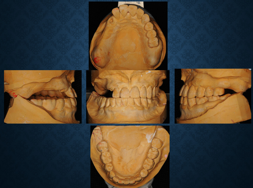
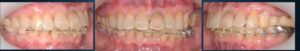
術前
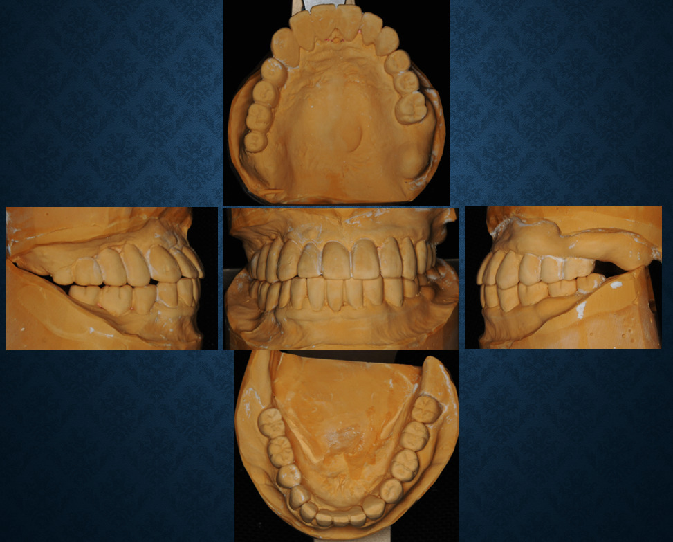
術後
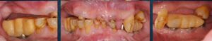
術前
術後


コメント