This is a case of periodontal treatment. A 60 years old female had chief complaints which were missing teeth and periodontal problems in remaining teeth, where original bone which hemmed in the teeth had disappeared because of gum disease bacteria. Implants placement for missing teeth and tissue regeneration surgery for the teeth which lost their surrounding bone were carried out. In order to check the results of the surgery, after 6 month, CT scanning was taken, which showed new bone regeneration to some extent, not perfect recovery. Incision, debridement, and suturing techniques must be more improved to get better results. Material choice may be also important. Growth mindset has to keep in mind and fixed mindset should be avoided in my learning curve.
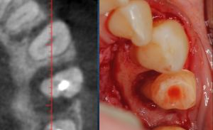
A CT horizontal section of the lower premolar whose surrounding bone was partially lost (right) a bird’s eye view of the site during the surgery (left)
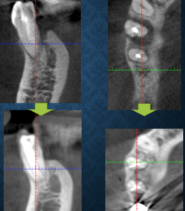
Vertical sections (left) and horizontal sections (right) of CT, before (above) and 6 months later (below)
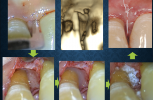
Photos during the surgery in the upper front tooth. Incision (above right), exposing defect (below left), applying Emdogain (below middle) , placing bone material (below left), suturing (above right) , 3D image before surgery (above middle)
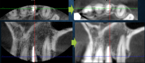
before (left) and 6 month (right) after surgery, horizontal (above) and vertical (below) sections


コメント